Classifying neurons for a better understanding of Stephen Hawking's disease
Understanding neurodegenerative diseases is a constant challenge for medical research. Amyotrophic lateral sclerosis (ALS), which affected famous physicist Stephen Hawking, is among these diseases. This pathology is characterized by the progressive death of so-called motor neurons that control muscles and that leading to paralysis. Understanding the processes responsible for the death of these neurons relies on the study of thousands of parameters that together define the healthy or sick status of a cell. In this context, artificial intelligence is a powerful tool that allows to quickly shed light on biological processes by simultaneously analyzing thousands of measurements, which is impossible for a human.
A database with more than 150,000 images
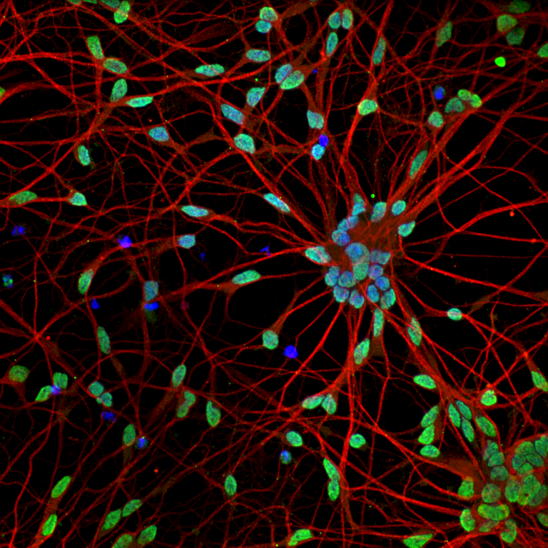
To understand what happens to neurons, you need data, tremendous amounts of data. Rickie Patani and Jasmine Harley from the Francis Crick Institute in London, specialized in medical research, have built a unique database of more than 150,000 images of motor neurons. These images were made using fluorescence microscopy. Thanks to a set of colors, this technique allows to distinguish different parts of the neurons, such as the nucleus and the dendrites. The cells were also subjected to different stress conditions, for example by inducing a thermal shock. Indeed, it has already been shown that under certain stress conditions, a healthy cell can look like a cell affected by ALS.

Intelligent classification to recognize affected neurons
This database was used to set up a model developed at Idiap and using artificial intelligence based on the technique known as deep learning. This model can distinguish whether a motor neuron is affected or not, thanks to the analysis of factors related to different parts of the cell. "To be effective, the model must also include enough different information in the set of images provided," Colombine Verzat, development engineer at Idiap and co-author of this publication, explains.
Unlike previous methods, this new model allows to analyse directly images pixel by pixel, without having to focus on a part of the cell or retain only a specific feature. This way the method is unbiased. "Tools offered by artificial intelligence are exciting because they allow us to grasp huge masses of data and to extract key information–tasks that surpass the human brain," Raphaëlle Luisier, head of the Genomics & health informatics research group, explains.
Guiding future research
The idea is to leverage the power of large image databases to test quickly hypotheses about how a disease works. For example, today, experts who study ALS know that mostly certain proteins in the cell are indicators of whether a neuron is affected. But this study shows that another part of motor neurons, the neurites, are also affected by ALS. This could guide further research into the influence of this cell structure in the disease. The ultimate goal is to improve the knowledge of this disease and to facilitate the discovery of a treatment, which does not exist to date.
More information
- Genomics & health informatics research group
